The strain first discovered at the end of November appears to be less severe and even patients who do end up in the hospital spend less time there, John Bell, regius professor of medicine at Oxford, said on BBC Radio 4’s Today program.
The omicron variant that’s taking the world by storm is not “the same disease we were seeing a year ago,” a University of Oxford immunologist said, reinforcing reports about the strain’s milder nature.
The strain first discovered at the end of November appears to be less severe and even patients who do end up in the hospital spend less time there, John Bell, regius professor of medicine at Oxford, said on BBC Radio 4’s Today program.
“The horrific scenes that we saw a year ago — intensive care units being full, lots of people dying prematurely — that is now history in my view, and I think we should be reassured that that’s likely to continue,” Bell said.
Bell’s comments came after the U.K. government said it wouldn’t introduce stricter Covid-19 restrictions in England before the end of the year.
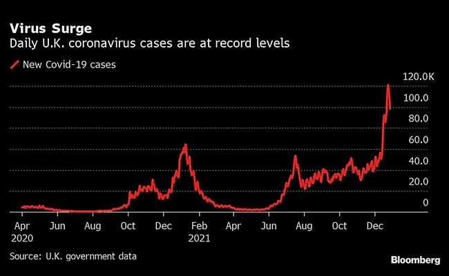
Infections have jumped by more than a quarter of a million in the past week, heaping pressure on Prime Minister Boris Johnson to respond. Health Secretary Sajid Javid late Monday said he’s monitoring the latest data and urged people to be careful, particularly at New Year celebrations.
A 30-year-old woman from the city of Esperanza, Argentina — the so-called Esperanza Patient — appears to be the second person whose immune system cleared the HIV-1 virus without antiretroviral therapy.
“During infection, HIV places copies of its genome into the DNA of cells, creating what is known as a viral reservoir,” said senior co-author Dr. Xu Yu, a researcher at Ragon Institute of MGH, MIT and Harvard Brigham and Women’s Hospital, and her colleagues.
“In this state, the virus effectively hides from anti-HIV drugs and the body’s immune response.”
“In most people, new viral particles are constantly made from this reservoir.”
“Antiretroviral therapy can prevent the new viruses from being made but cannot eliminate the reservoir, necessitating daily treatment to suppress the virus.”
“Some people, known as elite controllers, have immune systems that are able to suppress HIV without the need for medication.”
“Though they still have viral reservoirs that can produce more HIV virus, a type of immune cell called a killer T cell keeps the virus suppressed without the need for medication.”
In 2020, Dr. Yu and co-authors identified the first elite controller who had no intact HIV-1 viral sequence in her genome, indicating that her immune system may have eliminated the HIV-1 reservoir — what the scientists call a sterilizing cure.
The researchers sequenced billions of cells from that patient — known as the San Francisco Patient — searching for any HIV-1 sequence that could be used to create new virus, and found none.
The newly-identified patient, like the San Francisco Patient, has no intact HIV-1 genomes in a total of 1.188 billion peripheral blood mononuclear cells and 503 million mononuclear cells from placental tissues.
“These findings, especially with the identification of a second case, indicate there may be an actionable path to a sterilizing cure for people who are not able to do this on their own,” Dr. Yu said.
“The results may suggest a specific killer T cell response common to both patients driving this response, with the possibility that other people with HIV have also achieved a sterilizing cure.”
“If the immune mechanisms underlying this response can be understood by researchers, they may be able to develop treatments that teach others’ immune systems to mimic these responses in cases of HIV infection.”
“We are now looking toward the possibility of inducing this kind of immunity in persons on antiretroviral therapy through vaccination, with the goal of educating their immune systems to be able to control the virus without antiretroviral therapy,” she said.
Hope this seemingly magical recovery opens the doors to the ultimate cure/prevention for HIV infection!
There are around 20,000 of these proteins expressed by the human genome. Collectively, biologists refer to this full complement as the “proteome”.
Commenting on the results from AlphaFold, Dr Demis Hassabis, chief executive and co-founder of artificial intelligence company Deep Mind, said: “We believe it’s the most complete and accurate picture of the human proteome to date.
“We believe this work represents the most significant contribution AI has made to advancing the state of scientific knowledge to date.
“And I think it’s a great illustration and example of the kind of benefits AI can bring to society.” He added: “We’re just so excited to see what the community is going to do with this.”
Proteins are made up of chains of smaller building blocks called amino acids. These chains fold in myriad different ways, forming a unique 3D shape. A protein’s shape determines its function in the human body.
The 350,000 protein structures predicted by AlphaFold include not only the 20,000 contained in the human proteome, but also those of so-called model organisms used in scientific research, such as E. coli, yeast, the fruit fly and the mouse.
This giant leap in capability is described by DeepMind researchers and a team from the European Molecular Biology Laboratory (EMBL) in the prestigious journal Nature.
AlphaFold was able to make a confident prediction of the structural positions for 58% of the amino acids in the human proteome.
The positions of 35.7% were predicted with a very high degree of confidence – double the number confirmed by experiments.
Traditional techniques to work out protein structures include X-ray crystallography, cryogenic electron microscopy (Cryo-EM) and others. But none of these is easy to do: “It takes a huge amount of money and resources to do structures,” Prof John McGeehan, a structural biologist at the University of Portsmouth, told BBC News.
Therefore, the 3D shapes are often determined as part of targeted scientific investigations, but no project until now had systematically determined structures for all the proteins made by the body.
In fact, just 17% of the proteome is covered by a structure confirmed experimentally.
Commenting on the predictions from AlphaFold, Prof McGeehan said: “It’s just the speed – the fact that it was taking us six months per structure and now it takes a couple of minutes. We couldn’t really have predicted that would happen so fast.”
“When we first sent our seven sequences to the DeepMind team, two of those we already had the experimental structures for. So we were able to test those when they came back. It was one of those moments – to be honest – where the hairs stood up on the back of my neck because the structures [AlphaFold] produced were identical.”
Prof Edith Heard, from EMBL, said: “This will be transformative for our understanding of how life works. That’s because proteins represent the fundamental building blocks from which living organisms are made.”
“The applications are limited only by our understanding.”
Those applications we can envisage now include developing new drugs and treatments for disease, designing future crops that can resist climate change, and enzymes that can break down the plastic that pervades the environment.
Prof McGeehan’s group is already using AlphaFold’s data to help develop faster enzymes for degrading plastic. He said the program had provided predictions for proteins of interest whose structures could not be determined experimentally – helping accelerate their project by “multiple years”.
Dr Ewan Birney, director of EMBL’s European Bioinformatics Institute, said the AlphaFold predicted structures were “one of the most important datasets since the mapping of the human genome”.
DeepMind has teamed up with EMBL to make the AlphaFold code and protein structure predictions openly available to the global scientific community.
Dr Hassabis said DeepMind planned to vastly expand the coverage in the database to almost every sequenced protein known to science – over 100 million structures.
Chronic viral infections and cancer can cause “killer” T cells in the immune system to take on a state of dysfunction or exhaustion whereby they can no longer react to infectious invaders or abnormal cells like normal “memory” T cells. Two new studies led by investigators at Massachusetts General Hospital (MGH) and published in Nature Immunology provide insights into T cell exhaustion, which could lead to potential strategies to overcome it.
One study, which was led by Georg M. Lauer, MD, PhD, of the Division of Gastroenterology at MGH, focused on differences between memory and exhausted T cells in individuals with human hepatitis C virus (HCV) infection before and after treatment. After patients were treated and cured, their exhausted T cells tended to take on some properties of memory T cells but did not function as well as memory T cells.
“We saw some cosmetic improvement of the T cells that in a more superficial study could have been interpreted as real recovery, whereas in reality the key parameters determining the efficacy of a T cell were unchanged,” says Lauer. “A significant number of molecules that were altered were normalized after treatment, but others were stuck, and these were clearly the ones associated with T cell function.” This lack of recovery was especially prominent with a long duration of T cell stimulation by the virus; a shorter stimulation allowed the cells to revert to functional memory T cells.
“We are currently studying whether treating HCV with direct acting antiviral therapy in the acute phase of infection, instead of many years later, will result in full memory differentiation of T cells. If correct, this could indicate a short window of opportunity early during chronic infections to protect T cell function,” says Lauer.
Also, the molecules that the researchers found to be expressed in severely exhausted T cells might be targeted to rescue these cells.
A complementary study in the same issue of Nature Immunology that was led by Debattama Sen, PhD, at the Center for Cancer Research at MGH, and W. Nicholas Haining, BM, BCh, at Merck found that these exhausted T cells in chronic HCV infection were regulated epigenetically, or through physical changes in the cells’ chromosomes that affect the expression of genes. The investigators discovered that after clearing the virus, the epigenetic landscape of exhausted T cells was partially remodeled, but maintained many exhaustion-specific alterations, which the authors termed “epigenetic scars.” The epigenetic patterns paralleled the findings of the first paper on the protein and transcriptional level, indicating a key role for epigenetic control in determining the fate of the T cells. “These scars might be locking the exhausted T cells and preventing return to proper function even if the chronic infection in the patient is cured,” notes Sen. “Thus, restoring the function of these cells will likely require directly removing or inactivating these scarred regions to unlock the cells’ functionality.”
By comparing T cell responses across a range of viruses that are either effectively cleared (like influenza) or become chronic (like HCV and HIV), the scientists produced a map of where these exhaustion-specific scars occur. “This will enable precision editing and allow us to target the specific regions relevant to exhausted T cells and minimize off-target effects in other T cell populations,” says Sen.
The two studies were performed within an NIH/NIAID-funded U19 Cooperative Center on Human Immunology (CCHI) located at MGH. A third study on exhausted T cells, which was conducted by MGH CCHI investigators at the University of Pennsylvania, accompanies these two articles in Nature Immunology. A News & Views article in the journal provides additional perspectives on the implications of the studies’ findings.
exhausted immune cells in patients with chronic infections
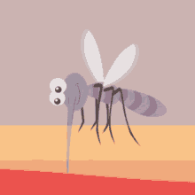
Monsoon can take a toll on human health. From manageable disease like cold and flu, to fatal diseases like dengue, malaria and chikungunya, monsoon brings along with it health complications that can put us at risk. While it might not be possible to avoid mosquito bites, as despite using ways like using mosquito repellents and avoiding mosquito-breeding, the vector succeeds in transmitting these diseases.
In a group, you must have noticed there is always someone who will complain about mosquitoes attacking them the most. That’s because, according to a report by Huff Post mosquitoes are selective insects, and some people are more likely to get bites than others.
There are certain factors which contribute to this effect. In one controlled study by the Journal of Medical Entomology, the bugs landed on people with blood Type O nearly twice as frequently as those with Type A. The researchers noted this has to do with secretions we produce, which tips mosquitoes off on a person’s blood type.
Entomology professor at the University of Florida, Jonathan F. Day said that more research needs to be conducted on mosquitoes’ potential preference for certain blood types over others. However, he agreed that mosquitoes do pick up on some cues we give off that make the bugs more likely to land on certain people.
“These cues let them know they are going to a blood source,” Day said. “Perhaps CO2 is the most important. The amount of CO2 you produce, like people with high metabolic rates ― genetic, other factors ― increases the amount of carbon dioxide you give off. The more you give off, the more attractive you are to these arthropods.”
The next question which pops up is what separates us from the nonliving entities that give off carbon dioxide, like cars? Mosquitoes look for primary cues in conjunction with what Day calls “secondary cues.”
Lactic acid — the stuff that causes our muscles to cramp during exercise — is one of those secondary cues, for example. Lactic acid is released through the skin, signaling to mosquitoes that we are a target, Day said.
Mosquitoes also have other qualities that help them pick up on secondary cues. “Mosquitoes have excellent vision, but they fly close to the ground to stay out of the wind,” Day said. “They are able to contrast you with the horizon, so how you’re dressed matters. If you have on dark clothes, you are going to attract more because you’ll stand out from the horizon, whereas those wearing light colors won’t as much.”
A mosquito also takes in “tactile cues” once it has landed on you.
“Body heat is a really important tactile cue,” Day said. “That comes into play with genetic differences or physiological differences. Some people tend to run a little warmer — when they land, they’re looking for a place where blood is close to the skin.” That means those whose temperatures are a little higher are more likely to get the bite.
Lifestyle or other health factors may also play a role, said Melissa Piliang, a dermatologist at Cleveland Clinic. “If body temperature is higher, you’re exercising and moving around a lot, or if you’re drinking alcohol, you are more attractive to mosquitoes,” Piliang said. “Being pregnant or being overweight also increases metabolic rate.”
Huff Post also said that one study showed that people who consumed just one can of beer were more at risk of attracting mosquitoes than those who didn’t. Of course, drinking outside is a popular summer and fall activity. “If you’ve been moving around all day doing yardwork and then you stop around dusk and drink a beer on your patio, you’re definitely at risk of bites,” Piliang said.
“Like a soldier or an athlete, innate immune cells can be trained by past experiences to become better at fighting infections.”
UCLA researchers have discovered the fundamental rule that allows the human body’s immune cells to be trained to aggressively respond to viruses, bacteria and other invaders, the university announced Thursday.
UCLA researchers identified a molecular mechanism within macrophages, which are infection-fighting cells in the innate immune system, that determines whether and how well the cells can be trained to fight invaders.
“Like a soldier or an athlete, innate immune cells can be trained by past experiences to become better at fighting infections,” said the study’s lead author, Quen Cheng, an assistant clinical professor of infectious diseases at UCLA’s Geffen School of Medicine.
Cheng noted that some experiences appear to be better than others for immune training, and that “this surprising finding motivated us to better understand the rules that govern this process.”
The study was published in the journal “Science” Friday, according to UCLA, which added that the findings could lead to strategies that enhance the immune system’s function.
Researchers found that immune training can occur if a cell’s DNA becomes unwrapped and exposes new genes that enable the cell to respond more aggressively, according to the study’s senior author Alexander Hoffman, professor of microbiology and director of the Institute for Quantitative and Computational Biosciences. When DNA is wrapped, only selected regions are exposed and accessible to fight an infection.
The UCLA researchers found that the precise dynamics of a key immune signaling molecule in macrophages, which is called NFKB and helps immune cells identify threats, determine if the DNA unwraps and genes are exposed.
Researchers also reported that the dynamic activity of NFKB itself is determined by the precise type of extracellular stimulus introduced to the macrophages.
“Importantly, our study shows that innate immune cells can be trained to become more aggressive only by some stimuli and not others,” Cheng said. “This specificity is critical to human health because proper training is important for effectively fighting infection, but improper training may result in too much inflammation and autoimmunity, which can cause significant damage.”
The NFKB is activated when receptors on the immune cells detect threatening external stimuli. The dynamics of NFKB form a language that UCLA researchers compared to Morse code — it communicates to the DNA that there is an external threat and tells the genes to get ready for battle.
Researchers used the bone marrow of mice to follow the activity of NFKB in macrophages, according to UCLA. They tracked how the molecule’s dynamics changed in response to several stimuli. NFKB was successful only when the stimulus induced non-oscillating NFKB activity.
“For a long time, we’ve known intuitively that whether NFKB oscillates or not must be important, but have simply not been able to figure out how,” Cheng said. “These results are a real breakthrough for understanding the language of immune cells, and knowing the language will help us `hack’ the system to improve immune function.”
The training process was simulated with a mathematical model, as well, UCLA said. Mathematical modeling of immune regulatory systems is a key goal of Hoffman’s laboratory.
Hoffman and Cheng expect to inspire a wide range of other studies from their research, including investigations into diseases caused by immune cells, strategies to improve immune training to fight infections and how to complement existing vaccine approaches.
“This study shows how collaborations between researchers in the UCLA College and David Geffen School of Medicine can produce innovative and impactful science that benefits human health,” Hoffmann said. Cheng earned his Ph.D. under Hoffman’s guidance at UCLA’s Specialty Training and Advanced Research program.
The study’s co-lead author is Sho Ohta, an assistant professor at the University of Tokyo and a former postdoctoral scholar in Hoffmann’s UCLA laboratory. Co-authors also include UCLA M.D. and Ph.D. student Katherine Sheu; Roberto Spreafico, a former postdoctoral scholar in Hoffmann’s laboratory; Adewunmi Adelaja, UCLA M.D. student who earned his Ph.D. in Hoffmann’s laboratory; and Brooks Taylor, a former UCLA doctoral student in Hoffmann’s laboratory.
The study was funded by UCLA’s Department of Medicine’s STAR Program and the National Institutes of Health.Copyright CNS – City News Service
I was watching Netflix series- “Alexa & Katie” which is about the high school journey two best friends one of whom suffers from leukemia (blood cancer). Watching it really motivated me to find out more about the life threatening disease- cancer which kills many people every year.
While surfing I came across this article about a novel research that optimizes that our body’s own immune system can fight cancer.
Before jumping on to the article, first, let’s look at what is cancer?
Cancer is a disease in which some of the body’s cells grow uncontrollably and spread to other parts of the body.
Cancer can start almost anywhere in the human body, which is made up of trillions of cells. Normally, human cells grow and multiply (through a process called cell division) to form new cells as the body needs them. When cells grow old or become damaged, they die, and new cells take their place.
Sometimes this orderly process breaks down, and abnormal or damaged cells grow and multiply when they shouldn’t. These cells may form tumors, which are lumps of tissue. Tumors can be cancerous.
Cancerous tumors spread into, or invade, nearby tissues and can travel to distant places in the body to form new tumors (a process called metastasis). Cancerous tumors may also be called malignant tumors. Many cancers form solid tumors, but cancers of the blood, such as leukemias, generally do not.
Armed with the basics of cancer let’s move on to the research article,
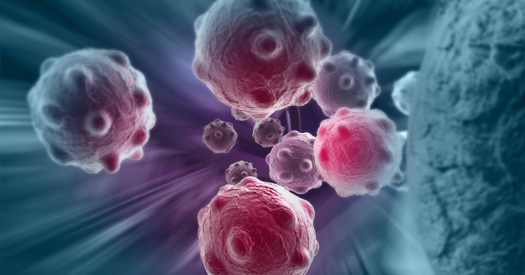
A groundbreaking study led by engineering and medical researchers at the University of Minnesota Twin Cities shows how engineered immune cells used in new cancer therapies can overcome physical barriers to allow a patient’s own immune system to fight tumors. The research could improve cancer therapies in the future for millions of people worldwide.
The research is published in Nature Communications, a peer-reviewed, open access, scientific journal published by Nature Research.
Instead of using chemicals or radiation, immunotherapy is a type of cancer treatment that helps the patient’s immune system fight cancer. T cells are a type of white blood cell that are of key importance to the immune system. Cytotoxic T cells are like soldiers who search out and destroy the targeted invader cells.
While there has been success in using immunotherapy for some types of cancer in the blood or blood-producing organs, a T cell’s job is much more difficult in solid tumors.
“The tumor is sort of like an obstacle course, and the T cell has to run the gauntlet to reach the cancer cells,” said Paolo Provenzano, the senior author of the study and a biomedical engineering associate professor in the University of Minnesota College of Science and Engineering. “These T cells get into tumors, but they just can’t move around well, and they can’t go where they need to go before they run out of gas and are exhausted.”
In this first-of-its-kind study, the researchers are working to engineer the T cells and develop engineering design criteria to mechanically optimize the cells or make them more “fit” to overcome the barriers. If these immune cells can recognize and get to the cancer cells, then they can destroy the tumor.
In a fibrous mass of a tumor, the stiffness of the tumor causes immune cells to slow down about two-fold — almost like they are running in quicksand.
“This study is our first publication where we have identified some structural and signaling elements where we can tune these T cells to make them more effective cancer fighters,” said Provenzano, a researcher in the University of Minnesota Masonic Cancer Center. “Every ‘obstacle course’ within a tumor is slightly different, but there are some similarities. After engineering these immune cells, we found that they moved through the tumor almost twice as fast no matter what obstacles were in their way.”
To engineer cytotoxic T cells, the authors used advanced gene editing technologies (also called genome editing) to change the DNA of the T cells so they are better able to overcome the tumor’s barriers. The ultimate goal is to slow down the cancer cells and speed up the engineered immune cells. The researchers are working to create cells that are good at overcoming different kinds of barriers. When these cells are mixed together, the goal is for groups of immune cells to overcome all the different types of barriers to reach the cancer cells.
Provenzano said the next steps are to continue studying the mechanical properties of the cells to better understand how the immune cells and cancer cells interact. The researchers are currently studying engineered immune cells in rodents and in the future are planning clinical trials in humans.
While initial research has been focused on pancreatic cancer, Provenzano said the techniques they are developing could be used on many types of cancers.
“Using a cell engineering approach to fight cancer is a relatively new field,” Provenzano said. “It allows for a very personalized approach with applications for a wide array of cancers. We feel we are expanding a new line of research to look at how our own bodies can fight cancer. This could have a big impact in the future.”
Honestly, let’s hope that this approach of treating cancer is able to save lives of millions of people affected with cancer
Cells inside our body keep dividing regularly, as is the scheme of the nature. However, if the division fails to take place, the cells will eventually die, leading to the chance of developing age-related diseases as well as cancer.
There are stretches of Deoxy Ribonucleic Acid (DNA) called telomeres present at the ends of chromosomes like protective caps. During cell division, these telomeres become shorter which makes the productivity of the protective cap less effective. Hence, the telomeres need to be checked upon regularly and elongated because if these DNA components get too short, the cell will stop dividing and move towards cell aging.
Scientists have studied what helps the telomeres to function properly and have found that a RNA species called TElomeric Repeat-containing RNA (TERRA) helps to work like the maintenance mechanic for telomeres. These get recruited at sites where telomeres need regulation and send a signal indicating that the telomeres need to be elongated or repaired. Which form of a system sends TERRA to the chromosome end is not known.
TERRA are a type of molecules called the non-coding RNAs, which do not get translated into proteins but instead function as chromosomes’ structural components. To study how these were getting assigned to places and remaining there, scientists visualized TERRA molecules under a microscope and found that a short stretch of the ribonucleic acid (RNA) was instrumental to bring it to the telomeres.
Now once TERRA has reached its required location, several proteins regulate its association with telomeres. Here, a protein called RAD51 plays a crucial role. Scientists from Ecole Polytechnique Fédérale de Lausanne and Masaryk University found that RAD51 was helping TERRA stick to telomeric DNA to form a so-called RNA-DNA hybrid molecule.
This sort of hybrid molecule formation has been previously detected only in the case of DNA repair. To witness it taking place during telomere repair is revolutionary.
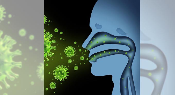
With a relatively minor genetic change, a new treatment developed by researchers at the Georgia Institute of Technology and Emory University appears to stop replication of both flu viruses and the virus that causes COVID-19. Best of all, the treatment could be delivered to the lungs via a nebulizer, making it easy for patients to administer themselves at home.
The therapy is based on a type of CRISPR, which normally allows researchers to target and edit specific portions of the genetic code, to target RNA molecules. In this case, the team used mRNA technology to code for a protein called Cas13a that destroys parts of the RNA genetic code that viruses use to replicate in cells in the lungs. It was developed by researchers in Philip Santangelo’s lab in the Wallace H. Coulter Department of Biomedical Engineering.
“In our drug, the only thing you have to change to go from one virus to another is the guide strand—we only have to change one sequence of RNA. That’s it,” Santangelo said. “We went from flu to SARS-CoV-2, the virus that causes COVID-19. They’re incredibly different viruses. And we were able to do that very, very rapidly by just changing a guide.”
The guide strand is a map that basically tells the Cas13a protein where to attach to the viruses’ RNA and begin to destroy it. Working with collaborators at the University of Georgia, Georgia State University, and Kennesaw State University, Santangelo’s team tested its approach against flu in mice and SARS-CoV-2 in hamsters. In both cases, the sick animals recovered.
Their results are reported Feb. 3 in the journal Nature Biotechnology. It’s the first study to show mRNA can be used to express the Cas13a protein and get it to work directly in lung tissue rather than in cells in a dish. It’s also the first to demonstrate the Cas13a protein is effective at stopping replication of SARS-CoV-2.
What’s more, the team’s approach has the potential to work against 99% of flu strains that have circulated over the last century. It also appears it would be effective against the new highly contagious variants of the coronavirus that have begun to circulate.
The key to that broad effectiveness is the sequence of genes the researchers target.
“In flu, we’re attacking the polymerase genes. Those are the enzymes that allow the virus to make more RNA and to replicate,” said Santangelo, the study’s corresponding author.
With help from a collaborator at the Centers for Disease Control and Prevention, they looked at the genetic sequences of prevalent flu strains over the last 100 years and found regions of RNA that are unchanged across nearly all of them.
“We went after those, because they’re far better conserved,” Santangelo said. “We let the biology dictate what our targets would be.”
Likewise, in SARS-CoV-2, the sequences the researchers targeted so far remain unchanged in the new variants.
The approach means the treatment is flexible and adaptable as new viruses emerge, said Daryll Vanover, a research scientist in Santangelo’s lab and the paper’s second author.
“One of the first things that society and the CDC is going to get when a pandemic emerges is the genetic sequence. It’s one of the first tools that the CDC and the surveillance teams are going to use to identify what kind of virus this is and to begin tracking it,” Vanover said. “Once the CDC publishes those sequences—that’s all we need. We can immediately screen across the regions that we’re interested in to target it and knock down the virus.”
Vanover said that can result in lead candidates for clinical trials in a matter of weeks—which is about how long it took them to scan the sequences, design their guide strands, and be ready for testing in this study.
“It’s really quite plug-and-play,” Santangelo said. “If you’re talking about small tweaks versus large tweaks, it’s a big bonus in terms of time. And in pandemics—if we had had a vaccine in a month or two after the pandemic hit, think about what things would look like now. If we had a therapy a month after it hit, what would things look like now? It could make a huge difference, the impact on the economy, the impact on people.”
The project was funded by the Defense Advanced Research Projects Agency’s (DARPA) PReemptive Expression of Protective Alleles and Response Elements (PREPARE) program, with the goal of creating safe, effective, transient, and reversible gene modulators as medical countermeasures that could be adapted and delivered rapidly. That’s why the team decided to try a nebulizer for delivering the treatment, Santangelo said.
“If you’re really trying to think of something that’s going to be a treatment that someone can actually give themselves in their own house, the nebulizer we used is not terribly different from one that you can go buy at a pharmacy,” he said.
The team’s approach also was sped along by their previous work on delivering mRNA to mucosal surfaces like those in the lungs. They knew there was a good chance they could tackle respiratory infections with that approach. They decided to use mRNA to code for the Cas13a protein because it’s an inherently safe technique.
“The mRNA is transient. It doesn’t get into the nucleus, doesn’t affect your DNA,” Santangelo said, “and for these CRISPR proteins, you really don’t want them expressed for long periods of time.”
He and Vanover said additional work remains—especially understanding more about the specific mechanisms that make the treatment effective. It has produced no side effects in the animal models, but they want to take a deeper look at safety as they consider moving closer to a therapy for human patients.
“This project really gave us the opportunity to push our limits in the lab in terms of techniques, in terms of new strategy,” said Chiara Zurla, the team’s project manager and a co-author on the paper. “Especially with the pandemic, we feel an obligation to do as much as we can as well as we can. This first paper is a great example, but many will follow; we’ve done a lot of work, and we have a lot of promising results.”
These sperm also take a straight path unlike their zigzagging rivals.
It turns out there’s a difference in competitiveness between sperm swimming towards an egg, and it’s down to genetics and one protein: RAC1.
If you thought it was by sheer luck that sperm make it all the way to the egg to fertilize it, you have just been proven wrong by a team of researchers at the Max Planck Institute for Molecular Genetics (MPIMG) in Berlin, Germany.
The researchers explained in a study, published in the journal PLOS Genetics on February 4, how a genetic factor called “t-haplotype” awards the success of reaching the egg first to sperm that contain it, and it happens 99 percent of the time.
In a first, the researchers pointed out that sperm with the t-haplotype moved faster than their peers without it. It also turned out that these faster-moving sperm were swimming straight, compared with their zigzagging competitors.
And it comes down to RAC1, a protein that transmits signals from the outside of the sperm cell to the inside by activating other proteins. It essentially helps to direct the sperm in the right direction.
IN DIRECT COMPETITION, T-SPERM WINS AGAINST THEIR NORMAL PEERS (+) IN THEIR RACE TO THE EGG USING GENETICS
On top of having assistance with directions, the t-haplotype sperm manage to poison their “normal” counterparts. These sperm not only produce a poison to stop their competitors, but they also create an antidote so that they themselves are protected from it, as Bernhard Herrmann, Director at the MPIMG and of the Institute of Medical Genetics at Charité – Universitätsmedizin Berlin, explained.
“Imagine a marathon, in which all participants get poisoned drinking water, but some runners also take an antidote,” Herrmann compares.
The team carried out its research on mice, so as to better understand the reasons for infertility in human males. Through their study, the researchers discovered that male mice with two copies of the t-haplotype were sterile because they produced only sperm with the t-haplotype, turning them all immotile. These cells have higher RAC1 levels.
However, having too low RAC1 levels also leads to disadvantages, as the sperm aren’t able to move quickly enough. So the researchers speculate that aberrant RAC1 activity may be the underlying reason for particular forms of male infertility.