Categories
Biology human body

Can body’s own immune system fight cancer?-turns out- YES!!

I was watching Netflix series- “Alexa & Katie” which is about the high school journey two best friends one of whom suffers from leukemia (blood cancer). Watching it really motivated me to find out more about the life threatening disease- cancer which kills many people every year.

While surfing I came across this article about a novel research that optimizes that our body’s own immune system can fight cancer.

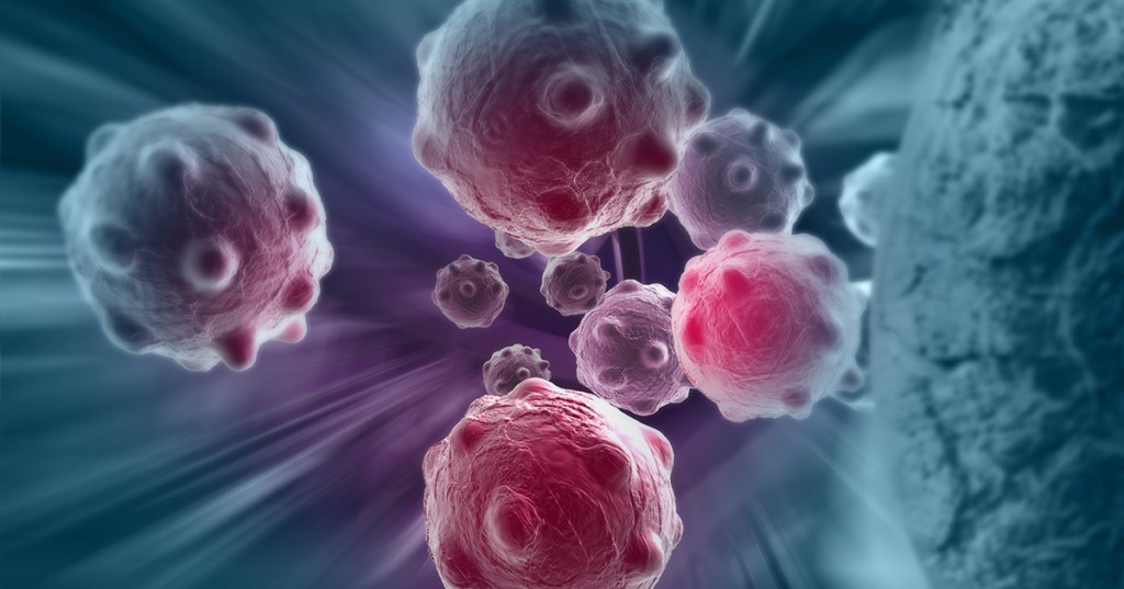
Before jumping on to the article, first, let’s look at what is cancer?

Cancer is a disease in which some of the body’s cells grow uncontrollably and spread to other parts of the body. 

Cancer can start almost anywhere in the human body, which is made up of trillions of cells. Normally, human cells grow and multiply (through a process called cell division) to form new cells as the body needs them. When cells grow old or become damaged, they die, and new cells take their place.

Sometimes this orderly process breaks down, and abnormal or damaged cells grow and multiply when they shouldn’t. These cells may form tumors, which are lumps of tissue. Tumors can be cancerous.

Cancerous tumors spread into, or invade, nearby tissues and can travel to distant places in the body to form new tumors (a process called metastasis). Cancerous tumors may also be called malignant tumors. Many cancers form solid tumors, but cancers of the blood, such as leukemias, generally do not.

Armed with the basics of cancer let’s move on to the research article,

A groundbreaking study led by engineering and medical researchers at the University of Minnesota Twin Cities shows how engineered immune cells used in new cancer therapies can overcome physical barriers to allow a patient’s own immune system to fight tumors. The research could improve cancer therapies in the future for millions of people worldwide.

The research is published in Nature Communications, a peer-reviewed, open access, scientific journal published by Nature Research.

Instead of using chemicals or radiation, immunotherapy is a type of cancer treatment that helps the patient’s immune system fight cancer. T cells are a type of white blood cell that are of key importance to the immune system. Cytotoxic T cells are like soldiers who search out and destroy the targeted invader cells.

While there has been success in using immunotherapy for some types of cancer in the blood or blood-producing organs, a T cell’s job is much more difficult in solid tumors.

“The tumor is sort of like an obstacle course, and the T cell has to run the gauntlet to reach the cancer cells,” said Paolo Provenzano, the senior author of the study and a biomedical engineering associate professor in the University of Minnesota College of Science and Engineering. “These T cells get into tumors, but they just can’t move around well, and they can’t go where they need to go before they run out of gas and are exhausted.”

In this first-of-its-kind study, the researchers are working to engineer the T cells and develop engineering design criteria to mechanically optimize the cells or make them more “fit” to overcome the barriers. If these immune cells can recognize and get to the cancer cells, then they can destroy the tumor.

In a fibrous mass of a tumor, the stiffness of the tumor causes immune cells to slow down about two-fold — almost like they are running in quicksand.

“This study is our first publication where we have identified some structural and signaling elements where we can tune these T cells to make them more effective cancer fighters,” said Provenzano, a researcher in the University of Minnesota Masonic Cancer Center. “Every ‘obstacle course’ within a tumor is slightly different, but there are some similarities. After engineering these immune cells, we found that they moved through the tumor almost twice as fast no matter what obstacles were in their way.”

To engineer cytotoxic T cells, the authors used advanced gene editing technologies (also called genome editing) to change the DNA of the T cells so they are better able to overcome the tumor’s barriers. The ultimate goal is to slow down the cancer cells and speed up the engineered immune cells. The researchers are working to create cells that are good at overcoming different kinds of barriers. When these cells are mixed together, the goal is for groups of immune cells to overcome all the different types of barriers to reach the cancer cells.

Provenzano said the next steps are to continue studying the mechanical properties of the cells to better understand how the immune cells and cancer cells interact. The researchers are currently studying engineered immune cells in rodents and in the future are planning clinical trials in humans.

While initial research has been focused on pancreatic cancer, Provenzano said the techniques they are developing could be used on many types of cancers.

“Using a cell engineering approach to fight cancer is a relatively new field,” Provenzano said. “It allows for a very personalized approach with applications for a wide array of cancers. We feel we are expanding a new line of research to look at how our own bodies can fight cancer. This could have a big impact in the future.”

Honestly, let’s hope that this approach of treating cancer is able to save lives of millions of people affected with cancer

Categories
Biology environment

Will your future clothes be made of algae?

Living materials, which are made by housing biological cells within a non-living matrix, have gained popularity in recent years as scientists recognize that often the most robust materials are those that mimic nature.

For the first time, an international team of researchers from the University of Rochester and Delft University of Technology in the Netherlands used 3D printers and a novel bioprinting technique to print algae into living, photosynthetic materials that are tough and resilient. The material has a variety of applications in the energy, medical, and fashion sectors. The research is published in the journal Advanced Functional Materials.

“Three-dimensional printing is a powerful technology for fabrication of living functional materials that have a huge potential in a wide range of environmental and human-based applications.” says Srikkanth Balasubramanian, a postdoctoral research associate at Delft and the first author of the paper. “We provide the first example of an engineered photosynthetic material that is physically robust enough to be deployed in real-life applications.”

HOW TO BUILD NEW MATERIALS: LIVING AND NONLIVING COMPONENTS??

To create the photosynthetic materials, the researchers began with a non-living bacterial cellulose — an organic compound that is produced and excreted by bacteria. Bacterial cellulose has many unique mechanical properties, including its flexibility, toughness, strength, and ability to retain its shape, even when twisted, crushed, or otherwise physically distorted.

The bacterial cellulose is like the paper in a printer, while living microalgae acts as the ink. The researchers used a 3D printer to deposit living algae onto the bacterial cellulose.

The combination of living (microalgae) and nonliving (bacterial cellulose) components resulted in a unique material that has the photosynthetic quality of the algae and the robustness of the bacterial cellulose; the material is tough and resilient while also eco-friendly, biodegradable, and simple and scalable to produce. The plant-like nature of the material means it can use photosynthesis to “feed” itself over periods of many weeks, and it is also able to be regenerated — a small sample of the material can be grown on-site to make more materials.

ARTIFICIAL LEAVES, PHOTOSYNTHETIC SKINS, AND BIO-GARMENTS

The unique characteristics of the material make it an ideal candidate for a variety of applications, including new products such as artificial leaves, photosynthetic skins, or photosynthetic bio-garments.

Artificial leaves are materials that mimic actual leaves in that they use sunlight to convert water and carbon dioxide — a major driver of climate change — into oxygen and energy, much like leaves during photosynthesis. The leaves store energy in chemical form as sugars, which can then be converted into fuels. Artificial leaves therefore offer a way to produce sustainable energy in places where plants don’t grow well, including outer space colonies. The artificial leaves produced by the researchers at Delft and Rochester are additionally made from eco-friendly materials, in contrast to most artificial leaf technologies currently in production, which are produced using toxic chemical methods.

“For artificial leaves, our materials are like taking the ‘best parts’ of plants — the leaves — which can create sustainable energy, without needing to use resources to produce parts of plants — the stems and the roots — that need resources but don’t produce energy,” says Anne S. Meyer, an associate professor of biology at Rochester. “We are making a material that is only focused on the sustainable production of energy.”

Another application of the material would be photosynthetic skins, which could be used for skin grafts, Meyer says. “The oxygen generated would help to kick-start healing of the damaged area, or it might be able to carry out light-activated wound healing.”

Besides offering sustainable energy and medical treatments, the materials could also change the fashion sector. Bio-garments made from algae would address some of the negative environmental effects of the current textile industry in that they would be high-quality fabrics that would be sustainability produced and completely biodegradable. They would also work to purify the air by removing carbon dioxide through photosynthesis and would not need to be washed as often as conventional garments, reducing water usage.

“Our living materials are promising because they can survive for several days with no water or nutrients access, and the material itself can be used as a seed to grow new living materials,” says Marie-Eve Aubin-Tam, an associate professor of bionanoscience at Delft. “This opens the door to applications in remote areas, even in space, where the material can be seeded on site.”

100% biodegradable T-shirt made from algae
100% biodegradable T-Shirt made from algae

Categories
Biology environment

New Technique Reliably Measures Whether Rivers or Lakes Have Run Out of Air

BOD and COD Measurement Methods

International study shows that freshwater polluted by fecal material can be determined more quickly and reliably using a new technique.

When wastewater from villages and cities flows into rivers and lakes, large quantities of fats, proteins, sugars and other carbon-containing, organic substances wind up in nature together with the fecal matter. These organic substances are broken down by bacteria that consume oxygen. The larger the volume of wastewater, the better the bacteria thrive. This, however, means the oxygen content of the water continues to decrease until finally the fish, mussels, or worms literally run out of air. This has created low-oxygen death zones in many rivers and lakes around the world.

No gold standard for measurements until now

In order to measure how heavily the waters are polluted with organic matter from feces, government bodies and environmental researchers regularly take water samples. One widely used measurement method uses a chemical reaction to determine the content of organic substances. As an international team of scientists now shows, this established method provides values from which the actual degree of the water pollution can hardly be derived. Prof. Helmuth Thomas, Director of Hereon’s Institute of Carbon Cycles is also a contributor to the study, which has now been published in the scientific journal Science Advances. “In the paper, we are therefore also introducing a new method for making the measurements much more reliable in the future,” he says.

Using the conventional measurement method, water samples are mixed with the chemicals permanganate or dichromate. These are especially reactive and break down all organic substances in a short time. The quantity of consumed permanganates or dichromates can then be used to determine how much organic substance was contained in the water sample.

Experts refer to this measurement as “chemical oxygen demand,” COD. The problem with the COD measurements is that they do not differentiate between the organic substances that wind up in the water with the sewage, and those that arise naturally — such as lignin and humic acids — which are released when wood decays. This means that the water pollution can hardly be distinguished from the natural content of organic substances.

“For the Han River in South Korea, for example, we have shown that the pollution with organic substances from wastewater in the past twenty-five years has decreased. The COD measurements, however, still show high values as they were before,” says Helmuth Thomas, “because here the natural substances make up a large portion of the organic matter in the water.”

Complicated biological analysis

But how can the actual pollution be measured more reliably? A biological measurement method has been established here for decades, but it is much more complex than the COD method and is therefore used more seldomly by government bodies and research institutions. In this case, a water sample is taken from the river or lake and the oxygen content of the water is measured as an initial value. Another “parallel sample” is immediately sealed airtight. Then this water sample rests for five days. During this time, the bacteria break down the organic substance, whereby they gradually consume the oxygen in the water. After five days, the container is opened and the oxygen is measured. If the water contains a great deal of organic matter, then the bacteria were particularly active. The oxygen consumption was then correspondingly high. Experts refer to the “biological oxygen demand” (BOD) in this measurement.

“The BOD measurement is far more precise than the COD because the bacteria preferentially break down the small organic molecules from the wastewater but leave the natural ones, such as lignin, untouched,” says Thomas. Nevertheless, the BOD measurement has its disadvantages, too. On the one hand, the BOD measurement takes five days, while the COD value is available after a few minutes. On the other, while filling, storing, and measuring the water samples, meticulous care must be taken to ensure that no oxygen from the ambient air winds up in the sample and falsifies the measurement value. “Only a few people with a great deal of laboratory experience have mastered how to entirely handle the BOD measurement,” says Thomas. “Therefore, government bodies and researchers even today still prefer the COD despite its greater uncertainties.”

Faster and more reliable method

Helmuth Thomas and his team are therefore introducing an alternative method that improves on the conventional BOD measurement. The advantage to the method is that only one water sample is necessary, which is immediately sealed and the oxygen consumption is measured without interfering with the sample. It is therefore unnecessary to open the sample after five days again to measure the oxygen content. This prevents the sample from coming into contact with atmospheric oxygen again.

With the new approach, an optical fiber is inserted into the sample vessel as soon as the water sample is filled. Through this fiber, the oxygen content can be continuously measured directly in the sample using optical effects. Thomas says, “We can measure the oxygen content non-stop and obtain a far more precise picture of the oxygen consumption by the bacteria.”

First tests have shown that a meaningful result is already available after about forty-eight hours, something that considerably accelerates the BOD measurement. All in all, the optical method makes the BOD measurements not only more reliable, but also faster. Helmuth Thomas assumes that the new method in the coming years therefore will be established as the new standard, which will replace both the COD as well as the classic BOD measurements. In the future, for example, it will be possible to determine more reliably than before whether water pollution control measures are actually successful.

Categories
Biology human body Uncategorized

Scientists Devise Technique to Stop Cells from Aging, Reduce Chances of Cancer

Cells inside our body keep dividing regularly, as is the scheme of the nature. However, if the division fails to take place, the cells will eventually die, leading to the chance of developing age-related diseases as well as cancer.

There are stretches of Deoxy Ribonucleic Acid (DNA) called telomeres present at the ends of chromosomes like protective caps. During cell division, these telomeres become shorter which makes the productivity of the protective cap less effective. Hence, the telomeres need to be checked upon regularly and elongated because if these DNA components get too short, the cell will stop dividing and move towards cell aging.

Scientists have studied what helps the telomeres to function properly and have found that a RNA species called TElomeric Repeat-containing RNA (TERRA) helps to work like the maintenance mechanic for telomeres. These get recruited at sites where telomeres need regulation and send a signal indicating that the telomeres need to be elongated or repaired. Which form of a system sends TERRA to the chromosome end is not known.

TERRA are a type of molecules called the non-coding RNAs, which do not get translated into proteins but instead function as chromosomes’ structural components. To study how these were getting assigned to places and remaining there, scientists visualized TERRA molecules under a microscope and found that a short stretch of the ribonucleic acid (RNA) was instrumental to bring it to the telomeres.

Now once TERRA has reached its required location, several proteins regulate its association with telomeres. Here, a protein called RAD51 plays a crucial role. Scientists from Ecole Polytechnique Fédérale de Lausanne and Masaryk University found that RAD51 was helping TERRA stick to telomeric DNA to form a so-called RNA-DNA hybrid molecule.

This sort of hybrid molecule formation has been previously detected only in the case of DNA repair. To witness it taking place during telomere repair is revolutionary.

FREEZE THE AGEING PROCESS
Categories
Biology Covid-19 human body

m-RNA treatment for flu and Covid-19 viruses

With a relatively minor genetic change, a new treatment developed by researchers at the Georgia Institute of Technology and Emory University appears to stop replication of both flu viruses and the virus that causes COVID-19. Best of all, the treatment could be delivered to the lungs via a nebulizer, making it easy for patients to administer themselves at home.

The therapy is based on a type of CRISPR, which normally allows researchers to target and edit specific portions of the genetic code, to target RNA molecules. In this case, the team used mRNA technology to code for a protein called Cas13a that destroys parts of the RNA genetic code that viruses use to replicate in cells in the lungs. It was developed by researchers in Philip Santangelo’s lab in the Wallace H. Coulter Department of Biomedical Engineering.

“In our drug, the only thing you have to change to go from one virus to another is the guide strand—we only have to change one sequence of RNA. That’s it,” Santangelo said. “We went from flu to SARS-CoV-2, the virus that causes COVID-19. They’re incredibly different viruses. And we were able to do that very, very rapidly by just changing a guide.”

The guide strand is a map that basically tells the Cas13a protein where to attach to the viruses’ RNA and begin to destroy it. Working with collaborators at the University of Georgia, Georgia State University, and Kennesaw State University, Santangelo’s team tested its approach against flu in mice and SARS-CoV-2 in hamsters. In both cases, the sick animals recovered.

Their results are reported Feb. 3 in the journal Nature Biotechnology. It’s the first study to show mRNA can be used to express the Cas13a protein and get it to work directly in lung tissue rather than in cells in a dish. It’s also the first to demonstrate the Cas13a protein is effective at stopping replication of SARS-CoV-2.

What’s more, the team’s approach has the potential to work against 99% of flu strains that have circulated over the last century. It also appears it would be effective against the new highly contagious variants of the coronavirus that have begun to circulate.

The key to that broad effectiveness is the sequence of genes the researchers target.

“In flu, we’re attacking the polymerase genes. Those are the enzymes that allow the virus to make more RNA and to replicate,” said Santangelo, the study’s corresponding author.

With help from a collaborator at the Centers for Disease Control and Prevention, they looked at the genetic sequences of prevalent flu strains over the last 100 years and found regions of RNA that are unchanged across nearly all of them.

“We went after those, because they’re far better conserved,” Santangelo said. “We let the biology dictate what our targets would be.”

Likewise, in SARS-CoV-2, the sequences the researchers targeted so far remain unchanged in the new variants.

The approach means the treatment is flexible and adaptable as new viruses emerge, said Daryll Vanover, a research scientist in Santangelo’s lab and the paper’s second author.

“One of the first things that society and the CDC is going to get when a pandemic emerges is the genetic sequence. It’s one of the first tools that the CDC and the surveillance teams are going to use to identify what kind of virus this is and to begin tracking it,” Vanover said. “Once the CDC publishes those sequences—that’s all we need. We can immediately screen across the regions that we’re interested in to target it and knock down the virus.”

Vanover said that can result in lead candidates for clinical trials in a matter of weeks—which is about how long it took them to scan the sequences, design their guide strands, and be ready for testing in this study.

“It’s really quite plug-and-play,” Santangelo said. “If you’re talking about small tweaks versus large tweaks, it’s a big bonus in terms of time. And in pandemics—if we had had a vaccine in a month or two after the pandemic hit, think about what things would look like now. If we had a therapy a month after it hit, what would things look like now? It could make a huge difference, the impact on the economy, the impact on people.”

The project was funded by the Defense Advanced Research Projects Agency’s (DARPA) PReemptive Expression of Protective Alleles and Response Elements (PREPARE) program, with the goal of creating safe, effective, transient, and reversible gene modulators as medical countermeasures that could be adapted and delivered rapidly. That’s why the team decided to try a nebulizer for delivering the treatment, Santangelo said.

“If you’re really trying to think of something that’s going to be a treatment that someone can actually give themselves in their own house, the nebulizer we used is not terribly different from one that you can go buy at a pharmacy,” he said.

The team’s approach also was sped along by their previous work on delivering mRNA to mucosal surfaces like those in the lungs. They knew there was a good chance they could tackle respiratory infections with that approach. They decided to use mRNA to code for the Cas13a protein because it’s an inherently safe technique.

“The mRNA is transient. It doesn’t get into the nucleus, doesn’t affect your DNA,” Santangelo said, “and for these CRISPR proteins, you really don’t want them expressed for long periods of time.”

He and Vanover said additional work remains—especially understanding more about the specific mechanisms that make the treatment effective. It has produced no side effects in the animal models, but they want to take a deeper look at safety as they consider moving closer to a therapy for human patients.

“This project really gave us the opportunity to push our limits in the lab in terms of techniques, in terms of new strategy,” said Chiara Zurla, the team’s project manager and a co-author on the paper. “Especially with the pandemic, we feel an obligation to do as much as we can as well as we can. This first paper is a great example, but many will follow; we’ve done a lot of work, and we have a lot of promising results.”

Categories
Biology human body

COMPETITIVE SPERMS SWIM FASTER AND POISON PEERS

These sperm also take a straight path unlike their zigzagging rivals.

It turns out there’s a difference in competitiveness between sperm swimming towards an egg, and it’s down to genetics and one protein: RAC1.

If you thought it was by sheer luck that sperm make it all the way to the egg to fertilize it, you have just been proven wrong by a team of researchers at the Max Planck Institute for Molecular Genetics (MPIMG) in Berlin, Germany. 

The researchers explained in a study, published in the journal PLOS Genetics on February 4, how a genetic factor called “t-haplotype” awards the success of reaching the egg first to sperm that contain it, and it happens 99 percent of the time. 

In a first, the researchers pointed out that sperm with the t-haplotype moved faster than their peers without it. It also turned out that these faster-moving sperm were swimming straight, compared with their zigzagging competitors. 

And it comes down to RAC1, a protein that transmits signals from the outside of the sperm cell to the inside by activating other proteins. It essentially helps to direct the sperm in the right direction.

IN DIRECT COMPETITION, T-SPERM WINS AGAINST THEIR NORMAL PEERS (+) IN THEIR RACE TO THE EGG USING GENETICS

On top of having assistance with directions, the t-haplotype sperm manage to poison their “normal” counterparts. These sperm not only produce a poison to stop their competitors, but they also create an antidote so that they themselves are protected from it, as Bernhard Herrmann, Director at the MPIMG and of the Institute of Medical Genetics at Charité – Universitätsmedizin Berlin, explained.

“Imagine a marathon, in which all participants get poisoned drinking water, but some runners also take an antidote,” Herrmann compares. 

The team carried out its research on mice, so as to better understand the reasons for infertility in human males. Through their study, the researchers discovered that male mice with two copies of the t-haplotype were sterile because they produced only sperm with the t-haplotype, turning them all immotile. These cells have higher RAC1 levels. 

However, having too low RAC1 levels also leads to disadvantages, as the sperm aren’t able to move quickly enough. So the researchers speculate that aberrant RAC1 activity may be the underlying reason for particular forms of male infertility.

Categories
Biology human body

BIOPRINTING : A giant leap in the healthcare system

Bioprinting is the medical and biotechnology equivalent of 3D printing. By using the same principles, the aim is to rapidly develop living structures resembling human-grown organs and tissue that can be used to heal people or test new drugs.

Precision Medicine

1088374106

Medical fields such as cosmetic surgery, tissue engineering, and regenerative medicine operate on the principle that we can precisely manufacture tissue or whole body parts to help the body repair itself. Bioprinting offers the accuracy of precisely measured biology alongside rapid manufacture so we can heal people more quickly and effectively.

Of course, printing biological tissue is much more complex than building a mechanical part. There are intricate layers of cells in living tissue that need growth factors and surrounding structures to develop successfully. To mimic this bioprinters use bioink made from cells, biochemical nutrients and biological scaffolds to support cells in a precise order.

Additional Materials Required

909024898

Bioinks have to operate under conditions that are suitable to living, growing tissue, so they cannot really be printed at temperatures that exceed body temperature. They use a binding agent – often a biopolymer gel, or hydrogel – that acts as the molecular scaffold.

Common components of bioinks include collagen and hyaluronic acid, active molecules found in extracellular matrix (ECM) in the body, plus substances like alginate, a vegetable biopolymer refined from seaweed that retains water. Creating the right cell matrix helps the cells to stick together, communicate with each other and turn into the right types of cells to make the target tissue.

The Process is Real

Perhaps the simplest form of bioprinting is inkjet printing. Bioink is sprayed through tiny nozzles so it has to be almost liquid and this limits the biological materials that can be printed. Most 3D printers operate by extruding material through a nozzle and bioprinters can use extrusion too, though care has to be taken not to damage cells through excessive force.

Other techniques such as laser-assisted bioprinting or electrospinning – which use laser and electrical energy to position cells – are incredibly precise and can be used with thicker bioinks, but they are more tricky to use with living cells and not as rapid or able to create large quantities of tissue.

Once the bioprinter has done its work then the post-processing stage begins. Bioreactor systems are often employed to help the tissue to mature. They can be used to mimic the forces and biochemical support that tissue needs to grow and differentiate correctly.

You Have So Much Potential

649528686

Bioprinting may be a relatively new field but the results so far are encouraging. Stem cells, which have the potential to turn into several types of cells, are being used to create bone or cartilage to mend fractures and joint injuries.

Australian researchers have used neural cells in a custom-made bioink to create a ‘benchtop brain‘ that allows medics to test brain function, new drugs and study brain disorders like schizophrenia. Meanwhile, regenerative medicine scientists in the US have created a bioprinter able to construct ear, muscle and bone structures with the right size, strength and function for implantation.

One of the primary goals of bioprinting is to create functioning internal organs such as livers, kidneys or hearts. By printing compatible organs using a patient’s own stem cells, the donor waiting list could become a thing of the past. To get to this point there have been some important breakthroughs in printing vascularized tissue in complex 3D

Organ printing can improve the health of society in general by eliminating the problem of diseases caused by organ failure, costly treatments and social care. That promise may be years away from realization but rapid prototyping enabled by bioprinting is pushing medical advances forward at pace.

Categories
Biology human body

CROWN OF NANOMEDICINE

An international team of researchers led by Michigan State University’s Morteza Mahmoudi has developed a new method to better understand how nanomedicines — emerging diagnostics and therapies that are very small yet very intricate — interact with patients’ biomolecules.

Medicines based on nanoscopic particles have the promise to be more effective than current therapies while reducing side effects. But subtle complexities have confined most of these particles to research labs and out of clinical use, said Mahmoudi, an assistant professor in the Department of Radiology and the Precision Health Program.

“There’s been a considerable investment of taxpayer money in cancer nanomedicine research, but that research hasn’t successfully translated to the clinic,” Mahmoudi said. “The biological effects of nanoparticles, how the body interacts with nanoparticles, remain poorly understood. And they need to be considered in detail.”

Mahmoudi’s team has now introduced a unique combination of microscopy techniques to enable more detailed consideration of those biological effects, which the researchers described in the journal Nature Communications, published online on Jan. 25.

The team’s methods let researchers see important differences between particles exposed to human plasma, the cell-free part of blood that contains biomolecules including proteins, enzymes and antibodies.

These biological bits latch onto a nanoparticle, creating a coating referred to as a corona (not to be confused with the novel coronavirus), the Latin word for crown. This corona contains clues about how nanoparticles interact with a patient’s biology. Now, Mahmoudi and his colleagues have shown how to get an unprecedented view of that corona.

“For the first time, we can image the 3-D structure of the particles coated with biomolecules at the nano level,” Mahmoudi said. “This is a useful approach to get helpful and robust data for nanomedicines, to get the kind of data that can affect scientists’ decisions about the safety and efficacy of nanoparticles.”

Although work like this is ultimately helping move therapeutic nanomedicines into the clinic, Mahmoudi is not optimistic that broad approval will happen any time soon. There’s still much to learn about the particles. Furthermore, one of the things that researchers do understand very well — that minute variations in these diminutive drugs can have outsized impact — was underscored by this study.

The researchers saw that the coronas of nanoparticles from the same batch, exposed to the same human plasma, could provoke a variety of reactions by a patient to a single dose.

Still, Mahmoudi sees an opportunity in this. He believes these particles could shine as diagnostics tools instead of drugs. Rather than trying to treat diseases with nanoscale medicine, he believes that persnickety particles would be well suited for the early detection of disease. For example, Mahmoudi’s group has previously shown this diagnostic potential for cancers and neurodegenerative diseases.

“We could become more proactive if we used nanoparticles as a diagnostic,” he said. “When you can detect disease at the earlier stages, it becomes easier to treat them.”

Nanomedicine | The Scientist Magazine®
NANOMEDICINE
Categories
Biology human body

THIRST: NEUROBIOLOGY

Neurobiology of Thirst: Neural Mechanisms that Control Hydration

Scientists at the Tokyo Institute of Technology (Tokyo Tech) provide deeper insights into neural thirst control. Their study published recently in Nature Communications indicates that cholecystokinin-mediated water-intake suppression is controlled by two neuronal ‘thirst-suppressing’ sub-populations in the subfornical organ in the brain; one population is persistently activated by excessive water levels, and the other, transiently after drinking water.

Water sustains life on earth. The first life originated in an ancient sea, and since then, nearly every species that has existed in the past or lives today depends on the exact balance of salt and water (~145 mM; called body-fluid homeostasis or salt homeostasis) for survival. Humans can go weeks without food but will not last more than a few days without water, stressing the importance of this liquid.

The human body has several intricate mechanisms to make sure we consume an appropriate amount of water for maintaining the homeostasis, which is requisite to survival. One of these simple but key “hacks,” is thirst. When the body experiences dehydration on a hot day (noted by the excess of sodium in the body compared to water, a condition called hypernatremia), the brain sends “signals” to the rest of the body, making us crave the tall glass of water. On the other hand, under a condition called hyponatremia, where there is a more water than sodium, we suppress water drinking. The neural mechanisms of how this happens are a subject of great interest.

A team of researchers from Tokyo Institute of Technology, headed by Prof Masaharu Noda, have conducted extensive research into this. In their previous studies, they identified that thirst is driven by the so-called “water neurons” in the subfornical organ (SFO) of the brain, a region just outside the blood-brain barrier. When the body is dehydrated, the plasma levels of a peptide hormone called angiotensin II increase. These levels are detected by special angiotensin II “receptors” of water neurons to stimulate water intake. In turn, under sodium-depleted conditions (where there is more water than sodium), the activity of these water neurons is suppressed by “GABAergic” interneurons. “The latter control appeared to be dependent on the hormone cholecystokinin (CCK) in the SFO. However, the CCK-mediated neural mechanisms underlying the inhibitory control of water intake had not been elucidated so far,” states Prof Noda.

NEUROBIOLOGY OF THIRST

Categories
Biology

WOO-HOO!! WE CAN NOW REVERSE CELL AGING!

Turning off a newly identified enzyme could reverse a natural aging process in cells.

Research findings by a KAIST team provide insight into the complex mechanism of cellular senescence and present a potential therapeutic strategy for reducing age-related diseases associated with the accumulation of senescent cells.

Simulations that model molecular interactions have identified an enzyme that could be targeted to reverse a natural aging process called cellular senescence. The findings were validated with laboratory experiments on skin cells and skin equivalent tissues, and published in the Proceedings of the National Academy of Sciences (PNAS). 

“Our research opens the door for a new generation that perceives aging as a reversible biological phenomenon,” says Professor Kwang-Hyun Cho of the Department of Bio and Brain engineering at the Korea Advanced Institute of Science and Technology (KAIST), who led the research with colleagues from KAIST and Amorepacific Corporation in Korea. 

Cells respond to a variety of factors, such as oxidative stress, DNA damage, and shortening of the telomeres capping the ends of chromosomes, by entering a stable and persistent exit from the cell cycle. This process, called cellular senescence, is important, as it prevents damaged cells from proliferating and turning into cancer cells. But it is also a natural process that contributes to aging and age-related diseases. Recent research has shown that cellular senescence can be reversed. But the laboratory approaches used thus far also impair tissue regeneration or have the potential to trigger malignant transformations. 

Professor Cho and his colleagues used an innovative strategy to identify molecules that could be targeted for reversing cellular senescence. The team pooled together information from the literature and databases about the molecular processes involved in cellular senescence. To this, they added results from their own research on the molecular processes involved in the proliferation, quiescence (a non-dividing cell that can re-enter the cell cycle) and senescence of skin fibroblasts, a cell type well known for repairing wounds. Using algorithms, they developed a model that simulates the interactions between these molecules. Their analyses allowed them to predict which molecules could be targeted to reverse cell senescence.

They then investigated one of the molecules, an enzyme called PDK1, in incubated senescent skin fibroblasts and three-dimensional skin equivalent tissue models. They found that blocking PDK1 led to the inhibition of two downstream signaling molecules, which in turn restored the cells’ ability to enter back into the cell cycle. Notably, the cells retained their capacity to regenerate wounded skin without proliferating in a way that could lead to malignant transformation.

The scientists recommend investigations are next done in organs and organisms to determine the full effect of PDK1 inhibition. Since the gene that codes for PDK1 is overexpressed in some cancers, the scientists expect that inhibiting it will have both anti-aging and anti-cancer effects.

THE SCIENTISTS CONDUCTED WHAT IS KNOWN AS AN ENSEMBLE MODEL SIMULATION TO IDENTIFY MOLECULES THAT COULD BE TARGETED TO REVERSE CELL SENESCENCE. THEY THEN USED THE MODEL TO PREDICT THE EFFECTS OF INHIBITING PDK1 IN SENESCENT CELLS, AND CONFIRMED THE RESULTS IN LAB-CULTURED CELLS AND SKIN EQUIVALENT TISSUE MODELS.
Design a site like this with WordPress.com
Get started